SRA-B308
Record
- Holding institution
- Institute of Dental History and Craniofacial Study, University of the Pacific
- Collection
- University of the Pacific (USA)
- Sex
- Male
Curator notes
CRA - right frontal and sphenoid; left maxilla; bilateral zygomatic arch; right nasal
Scientific notes
Several fractures: Incision; Transverse with Displacement; Depressed; Linear
Thrusting.
All fractures show signs of healing.
4 lesions
sharp
- Bones
- Cranium
- Side
- Right
- Timing
- Antemortem
- Force
- Direct
- Fracture type
- Torsion, Compression
Unknown Unknown
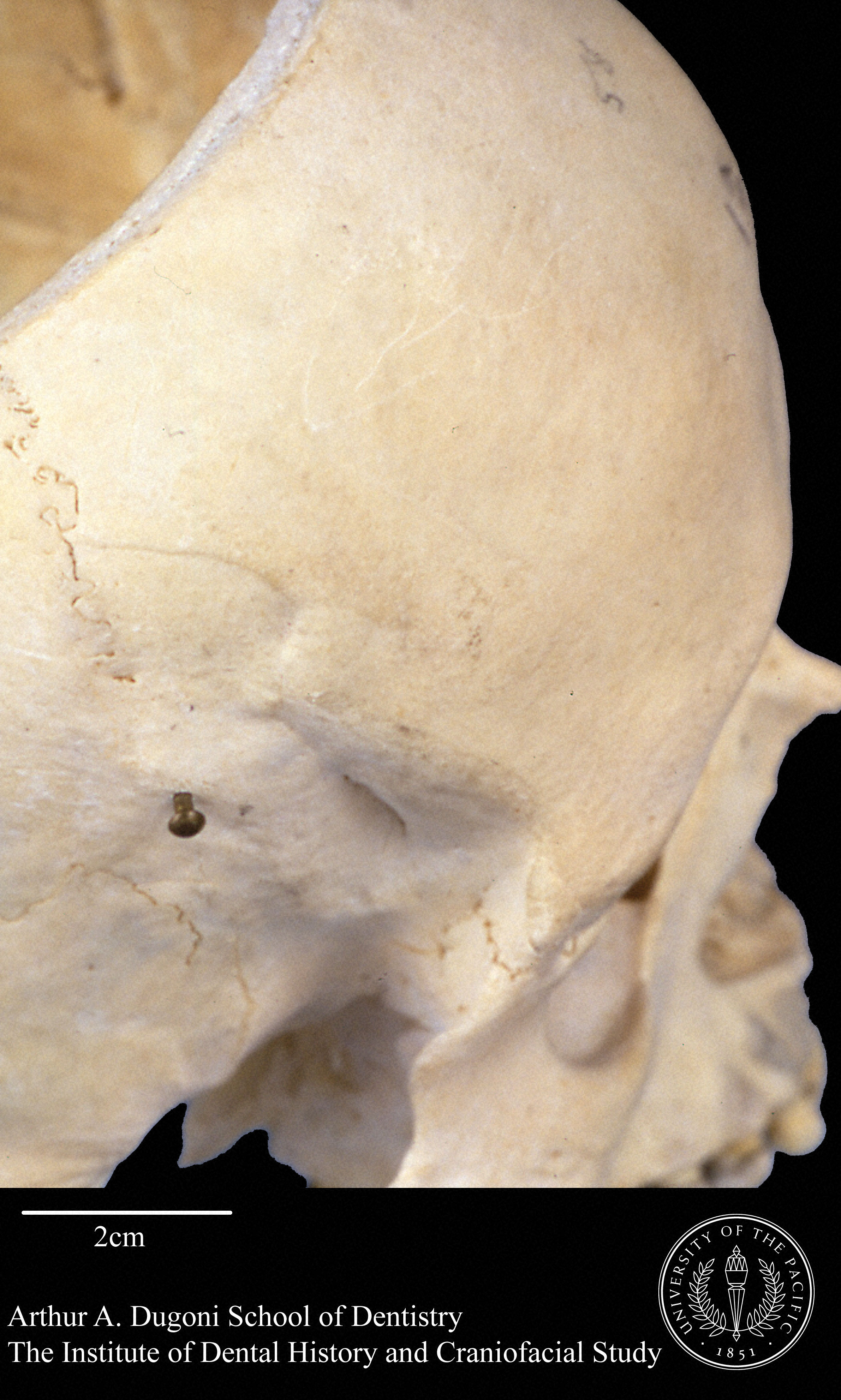
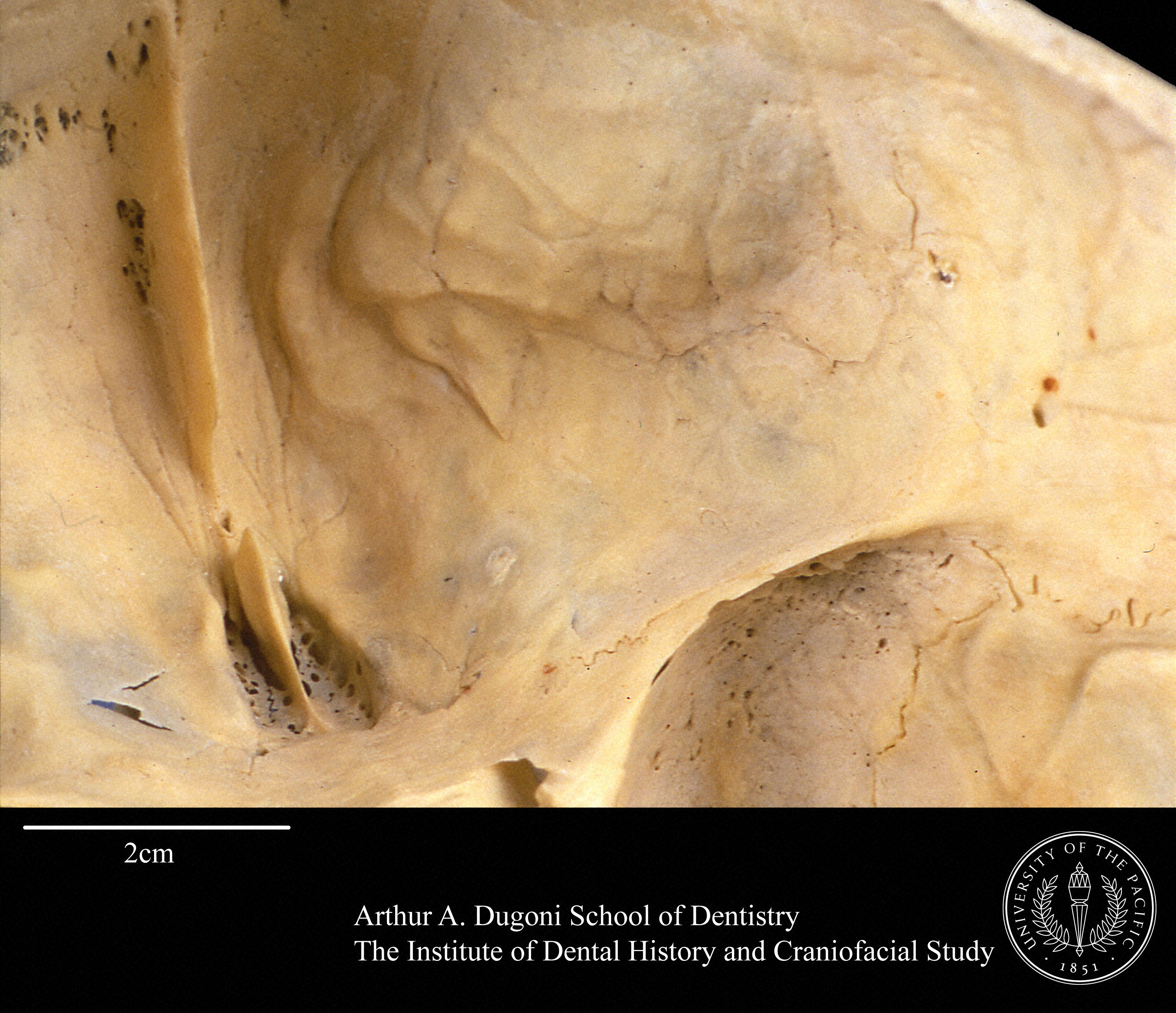
A sharp object lesion to the ectocranial surface of the right frontal has caused development of a dense bone callus, 1 cm thick and ~3x5cm in size, on the endocranial surface. The lesion suggests a posterior to anterior direction of thrust. The injury also involves the greater wing of the sphenoid.
-
contact: <a href="http://dental.pacific.edu/x1581.xml">Dorothy Dechant and Gary D. Richards</a> <br />Photo: Jon Draper <br /> license -> <a href="http://dental.pacific.edu/Institute_of_Dental_History_and_Craniofacial_Study/Museum_Collection_Policies/Images_of_Specimens_from_the_SRA_and_CA_Collections.html">Institute of Dental History and Craniofacial Study</a>
-
contact: <a href="http://dental.pacific.edu/x1581.xml">Dorothy Dechant and Gary D. Richards</a> <br />Photo: Jon Draper <br /> license -> <a href="http://dental.pacific.edu/Institute_of_Dental_History_and_Craniofacial_Study/Museum_Collection_Policies/Images_of_Specimens_from_the_SRA_and_CA_Collections.html">Institute of Dental History and Craniofacial Study</a>
sharp
- Bones
- Cranium
- Side
- Left
- Timing
- Antemortem
- Fracture type
- Compression, Tension
Unknown Unknown
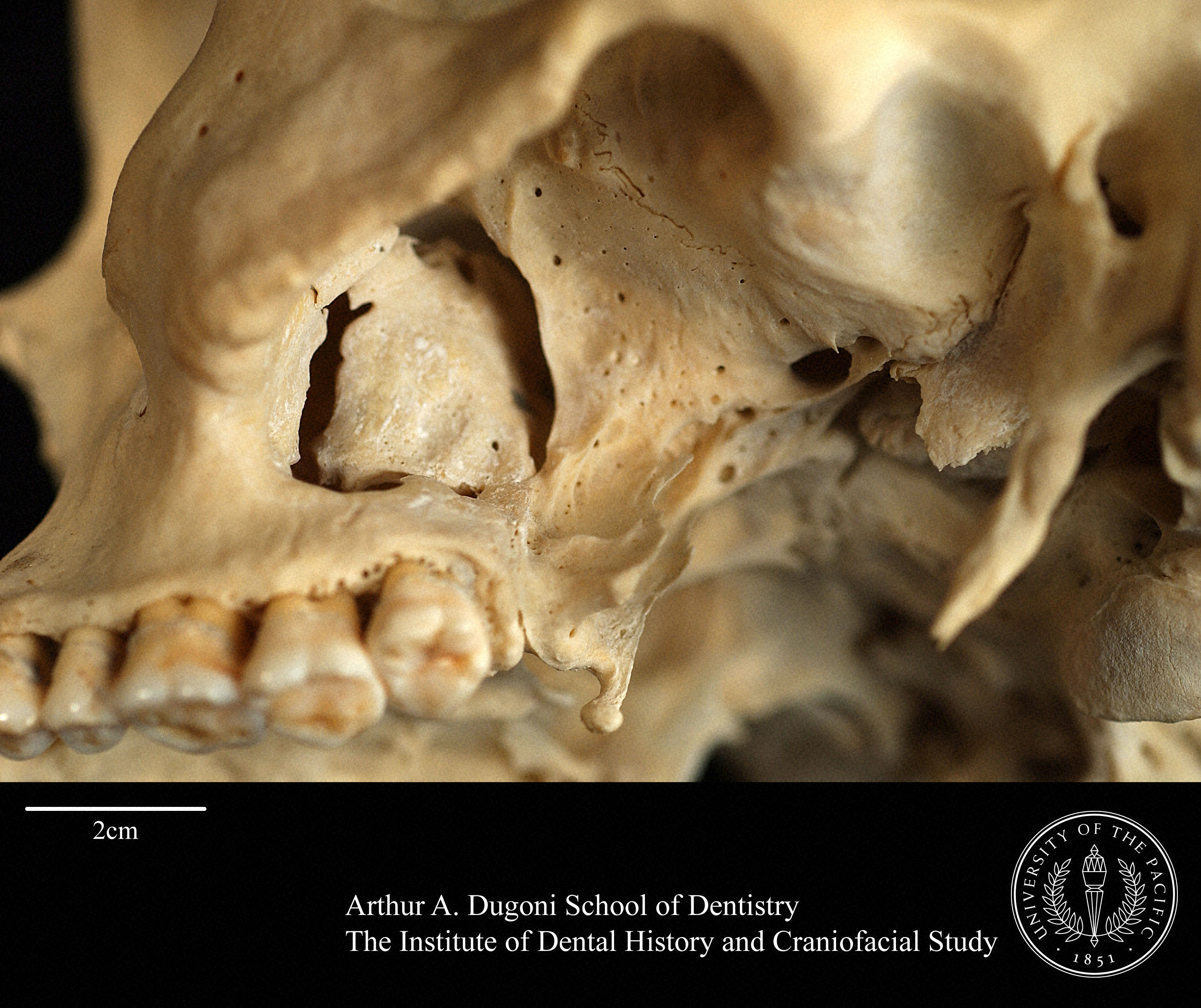
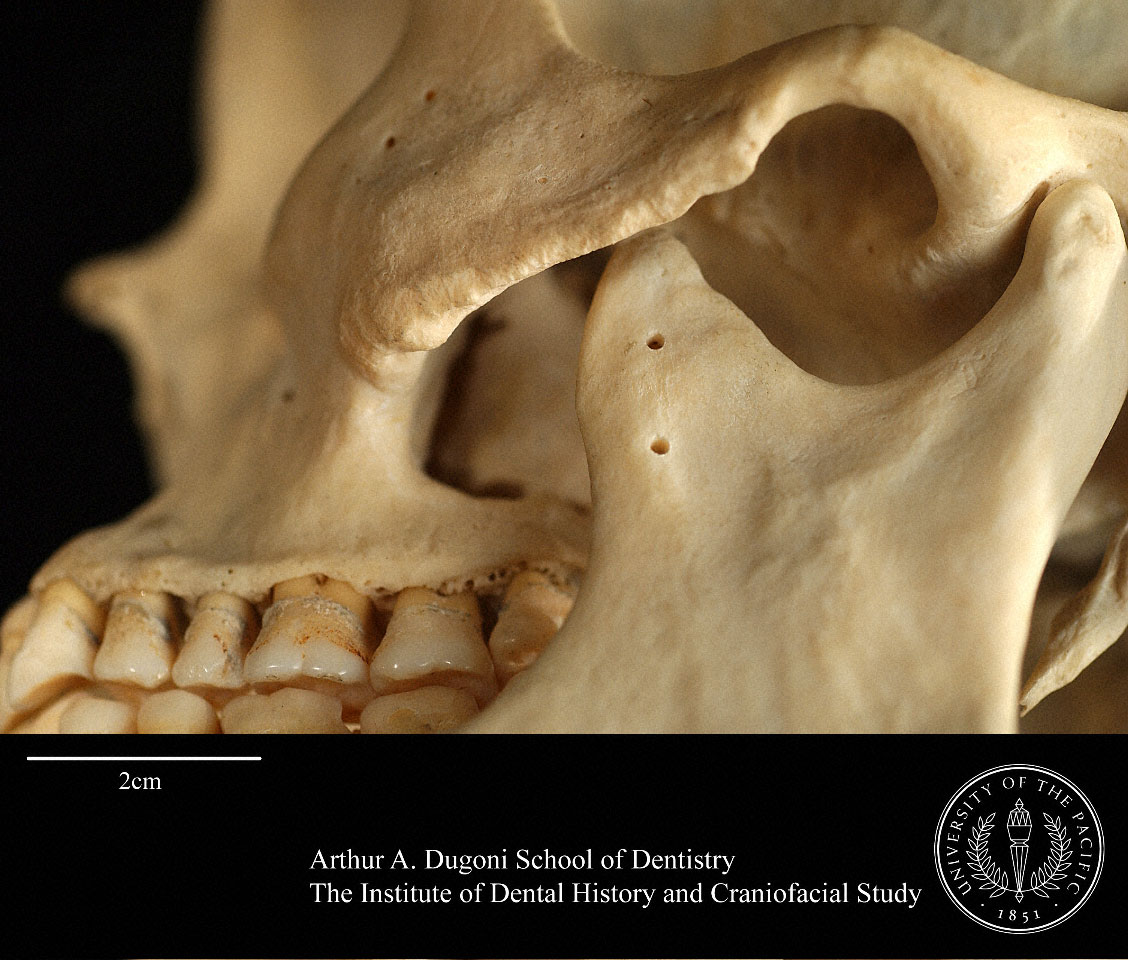
A second sharp object lesion has resulted in a depressed fracture to the lateral wall of the left maxillary sinus, with separation at the zygomaxillary suture and pterygoid plate. The resulting patent maxillary sinus suggests sharp object penetration passing just inferior to the zygomatic arch and anterior to the coronoid process of the mandible, driving the lateral maxillary wall superomedially. No damage to the mandible is evident.
-
contact: <a href="http://dental.pacific.edu/x1581.xml">Dorothy Dechant and Gary D. Richards</a> <br />Photo: Dorothy Dechant and Gary D. Richards <br /> licence -> <a href="http://dental.pacific.edu/Institute_of_Dental_History_and_Craniofacial_Study/Museum_Collection_Policies/Images_of_Specimens_from_the_SRA_and_CA_Collections.html">Institute of Dental History and Craniofacial Study</a>
-
contact: <a href="http://dental.pacific.edu/x1581.xml">Dorothy Dechant and Gary D. Richards</a> <br />Photo: Dorothy Dechant and Gary D. Richards <br /> license -> <a href="http://dental.pacific.edu/Institute_of_Dental_History_and_Craniofacial_Study/Museum_Collection_Policies/Images_of_Specimens_from_the_SRA_and_CA_Collections.html">Institute of Dental History and Craniofacial Study</a>
blunt
- Bones
- Cranium
- Timing
- Antemortem
- Fracture type
- Compression
Unknown Unknown
The right zygomatic process of the temporal exhibits a transverse fracture displaced medially with a linear fracture through a portion of the articular eminence.
-
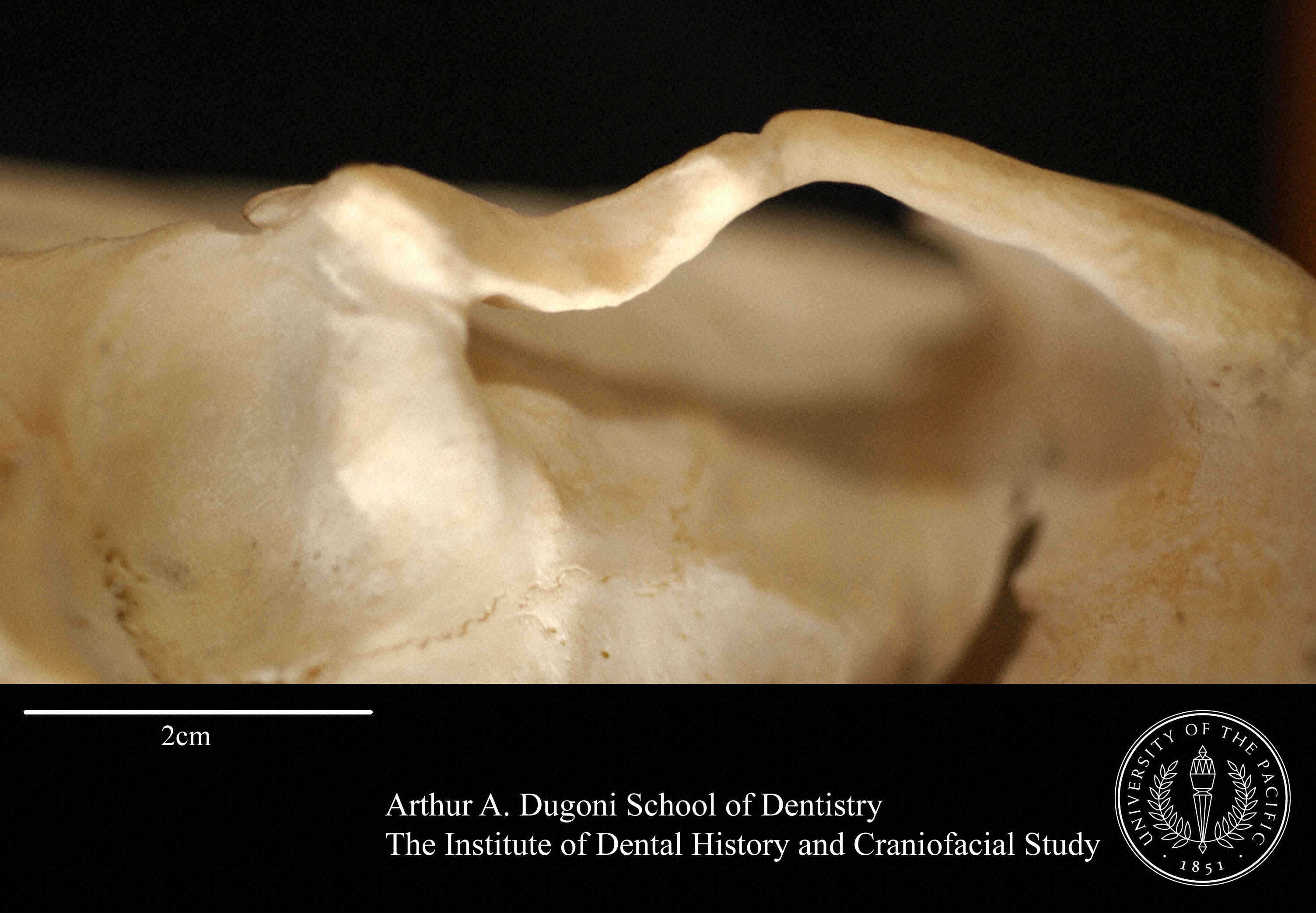
contact: <a href="http://dental.pacific.edu/x1581.xml">Dorothy Dechant and Gary D. Richards</a> <br />Photo: Dorothy Dechant and Gary D. Richards <br /> license -> <a href="http://dental.pacific.edu/Institute_of_Dental_History_and_Craniofacial_Study/Museum_Collection_Policies/Images_of_Specimens_from_the_SRA_and_CA_Collections.html">Institute of Dental History and Craniofacial Study</a>
blunt
- Bones
- Cranium
- Timing
- Antemortem
Unknown Unknown
The left zygomatic also shows a linear fracture through the articular eminence, but the left arch is less displaced than the right. The orbits appear unaffected. The palate shows some asymmetry in all directions that may have resulted from the traumas.
-
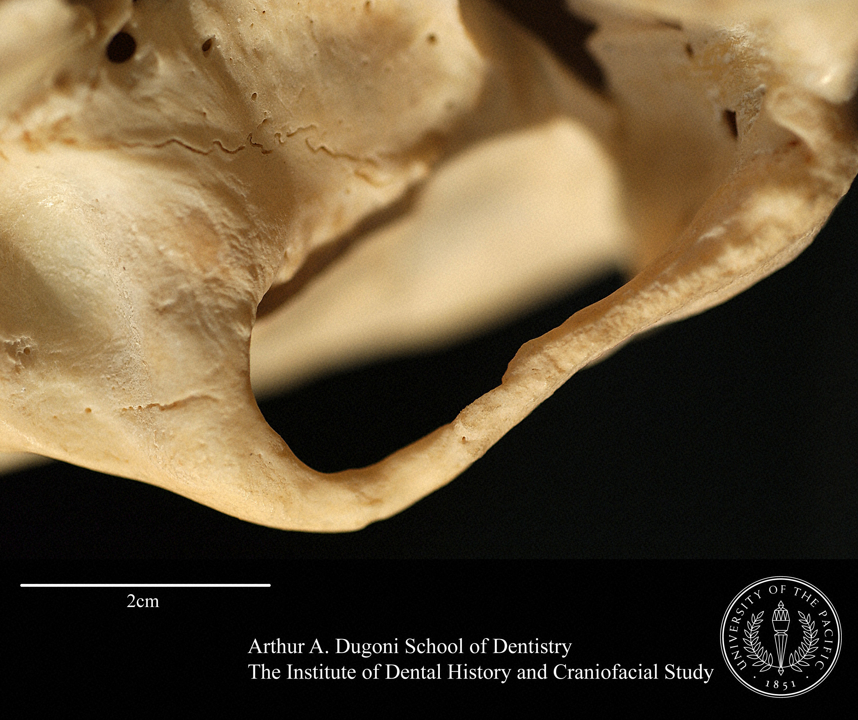
contact: <a href="http://dental.pacific.edu/x1581.xml">Dorothy Dechant and Gary D. Richards</a> <br />Photo: Dorothy Dechant and Gary D. Richards <br /> license -> <a href="http://dental.pacific.edu/Institute_of_Dental_History_and_Craniofacial_Study/Museum_Collection_Policies/Images_of_Specimens_from_the_SRA_and_CA_Collections.html">Institute of Dental History and Craniofacial Study</a>
-
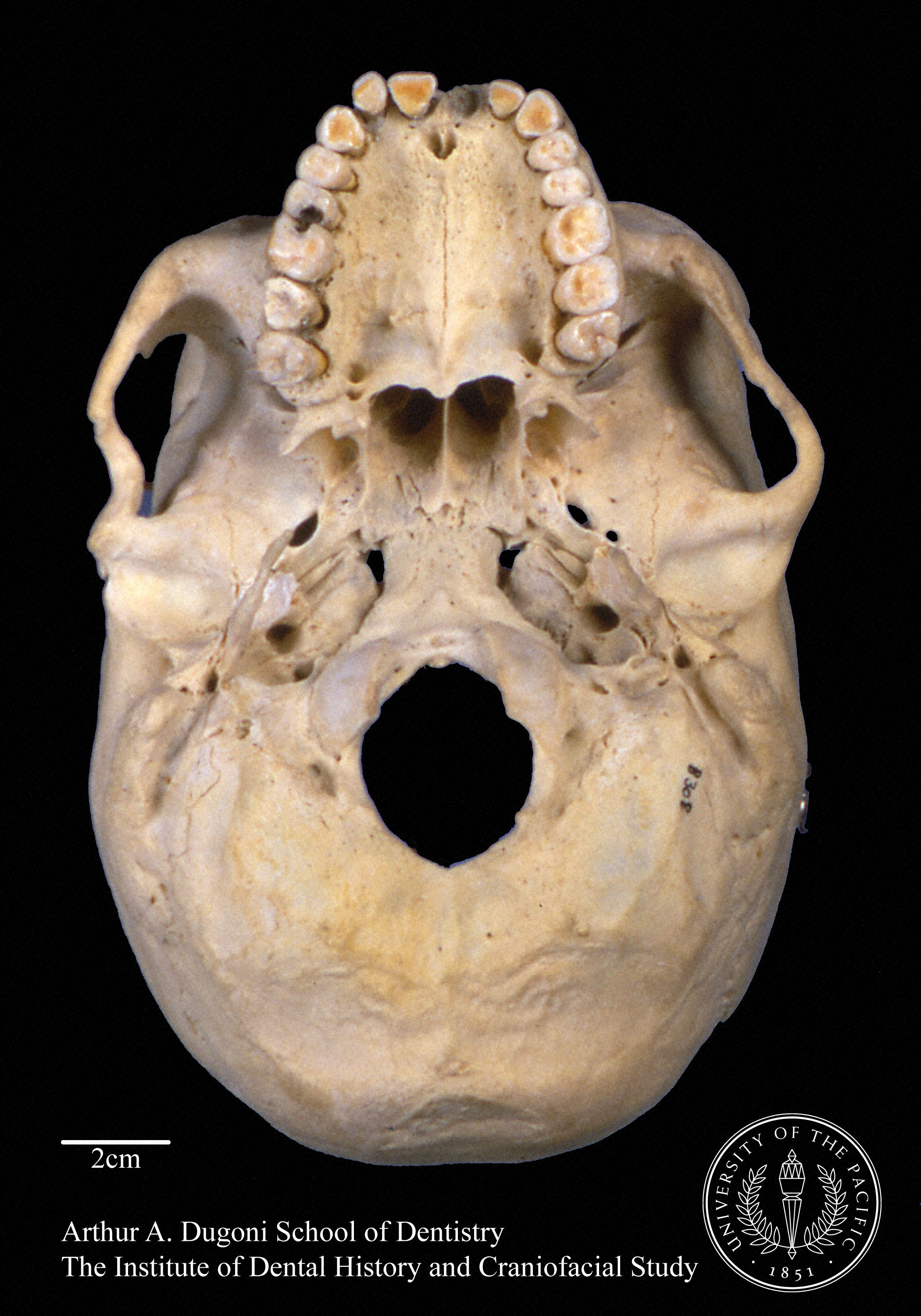
contact: <a href="http://dental.pacific.edu/x1581.xml">Dorothy Dechant and Gary D. Richards</a> <br />Photo: Jon Draper <br /> license -> <a href="http://dental.pacific.edu/Institute_of_Dental_History_and_Craniofacial_Study/Museum_Collection_Policies/Images_of_Specimens_from_the_SRA_and_CA_Collections.html">Institute of Dental History and Craniofacial Study</a>
Citing this record
Cite the holding institution. FOROST is a finding aid, not the rights holder.
Institute of Dental History and Craniofacial Study, University of the Pacific. University of the Pacific (USA). catalogue no. SRA-B308
Institute of Dental History and Craniofacial Study, University of the Pacific